Topic: Biopolymers. Nucleic acids, ATP and other organic compounds
Full title educational institution: Department general education Tomsk region Branch of the regional state educational institution "Tomsk State Pedagogical College" in Kolpashevo
Well: Biology
Chapter:General biology
Subject: Biopolymers. Nucleic acids, ATP and others organic compounds.
Purpose of the lesson: continue the study of biopolymers, contribute to the formation of logical techniques and cognitive abilities.
Lesson objectives:
Educational: introduce students to the concepts of nucleic acids, promote comprehension and assimilation of the material.
Educational: develop the cognitive qualities of students (the ability to see a problem, the ability to ask questions).
Educational: to form positive motivation for studying biology, the desire to obtain the final result, the ability to make decisions and draw conclusions.
Implementation time: 90 min.
Equipment:
· handout didactic material (list of amino acid coding);
Plan:
1. Types of nucleic acids.
2. Structure of DNA.
3. Main types of RNA.
4. Transcription.
5. ATP and other organic compounds of the cell.
Progress of the lesson:
I. Organizational moment.
Checking readiness for class.
II. Repetition.
Oral survey:
1. Describe the functions of fats in the cell.
2. What is the difference between protein biopolymers and carbohydrate biopolymers? What are their similarities?
Testing(3 options)
III. Learning new material.
1. Types of nucleic acids. The name nucleic acids comes from the Latin word nucleos, meaning nucleus: they were first discovered in cell nuclei. There are two types of nucleic acids in cells: deoxyribonucleic acid (DNA) and ribonucleic acid (RNA). These biopolymers are made up of monomers called nucleotides. The nucleotide monomers of DNA and RNA are similar in basic structural features and play a central role in storage and transmission hereditary information. Each nucleotide consists of three components connected by strong chemical bonds. Each of the nucleotides that make up RNA contains a tricarbon sugar - ribose; one of four organic compounds called nitrogenous bases - adenine, guanine, cytosine, uracil (A, G, C, U); phosphoric acid residue.
2. Structure of DNA . The nucleotides that make up DNA contain a five-carbon sugar - deoxyribose; one of four nitrogenous bases: adenine, guanine, cytosine, thymine (A, G, C, T); phosphoric acid residue.
In the composition of nucleotides, a nitrogenous base is attached to a molecule of ribose (or deoxyribose) on one side, and a phosphoric acid residue on the other. The nucleotides are connected to each other into long chains. The backbone of such a chain is formed by regularly alternating sugar and phosphoric acid residues, and the side groups of this chain are four type of irregularly alternating nitrogenous bases.
The DNA molecule is a structure consisting of two strands, which are connected to each other along their entire length by hydrogen bonds. This structure, unique to DNA molecules, is called a double helix. A feature of the DNA structure is that opposite the nitrogenous base A in one chain lies the nitrogenous base T in the other chain, and the nitrogenous base C is always located opposite the nitrogenous base G.
Schematically, what has been said can be expressed as follows:
A (adenine) - T (thymine)
T (thymine) - A (adenine)
G (guanine) - C (cytosine)
C (cytosine) - G (guanine)
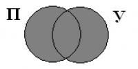
These pairs of bases are called complementary bases (complementing each other). DNA strands in which the bases are located complementary to each other are called complementary strands.
The model of the structure of the DNA molecule was proposed by J. Watson and F. Crick in 1953. It was fully confirmed experimentally and played an extremely important role in the development of molecular biology and genetics.
The order of arrangement of nucleotides in DNA molecules determines the order of arrangement of amino acids in linear protein molecules, i.e., their primary structure. A set of proteins (enzymes, hormones, etc.) determines the properties of the cell and the organism. DNA molecules store information about these properties and pass them on to generations of descendants, i.e. they are carriers of hereditary information. DNA molecules are mainly found in the nuclei of cells and in small quantities in mitochondria and chloroplasts.
3. Main types of RNA. Hereditary information stored in DNA molecules is realized through protein molecules. Information about the structure of the protein is transmitted to the cytoplasm by special RNA molecules, which are called messenger RNA (i-RNA). Messenger RNA is transferred to the cytoplasm, where protein synthesis occurs with the help of special organelles - ribosomes. It is messenger RNA, which is built complementary to one of the DNA strands, that determines the order of amino acids in protein molecules.
Another type of RNA also takes part in protein synthesis - transport RNA (t-RNA), which brings amino acids to the place of formation of protein molecules - ribosomes, a kind of factories for the production of proteins.
Ribosomes contain a third type of RNA, the so-called ribosomal RNA (r-RNA), which determines the structure and functioning of ribosomes.
Each RNA molecule, unlike a DNA molecule, is represented by a single strand; It contains ribose instead of deoxyribose and uracil instead of thymine.
So, Nucleic acids perform the most important biological functions in the cell. DNA stores hereditary information about all the properties of the cell and the organism as a whole. Different kinds RNAs take part in the implementation of hereditary information through protein synthesis.
4. Transcription.
The process of mRNA formation is called transcription (from the Latin “transcription” - rewriting). Transcription occurs in the cell nucleus. DNA → mRNA with the participation of the polymerase enzyme. t-RNA performs the function of a translator from the “language” of nucleotides to the “language” of amino acids, t-RNA receives a command from i-RNA - the anticodon recognizes the codon and carries the amino acid.
 The final product" href="/text/category/konechnij_produkt/" rel="bookmark">The final products of biosynthesis include amino acids, from which proteins are synthesized in cells; nucleotides - monomers, from which nucleic acids (RNA and DNA) are synthesized; glucose, which serves as a monomer for the synthesis of glycogen, starch, and cellulose.
The path to the synthesis of each of the final products lies through a series of intermediate compounds. Many substances undergo enzymatic breakdown and breakdown in cells.
The end products of biosynthesis are substances that play an important role in the regulation physiological processes and development of the body. These include many animal hormones. Hormones of anxiety or stress (for example, adrenaline) under conditions of tension increase the release of glucose into the blood, which ultimately leads to an increase in ATP synthesis and active use of energy stored by the body.
Adenosine phosphoric acids. A particularly important role in the bioenergetics of the cell is played by the adenyl nucleotide, to which two more phosphoric acid residues are attached. This substance is called adenosine triphosphoric acid (ATP). ATP molecule is a nucleotide formed nitrogenous base adenine, the five-carbon sugar ribose and three phosphoric acid residues. The phosphate groups in the ATP molecule are connected to each other by high-energy (macroergic) bonds.
ATP- universal biological energy accumulator. The light energy of the Sun and the energy contained in the food consumed are stored in ATP molecules.
The average lifespan of 1 ATP molecule in the human body is less than a minute, so it is broken down and restored 2400 times a day.
IN chemical bonds Energy (E) is stored between the phosphoric acid residues of the ATP molecule, which is released when the phosphate is removed:
ATP = ADP + P + E
This reaction produces adenosine diphosphoric acid (ADP) and phosphoric acid (phosphate, P).
ATP + H2O → ADP + H3PO4 + energy (40 kJ/mol)
ATP + H2O → AMP + H4P2O7 + energy (40 kJ/mol)
ADP + H3PO4 + energy (60 kJ/mol) → ATP + H2O
All cells use ATP energy for the processes of biosynthesis, movement, heat production, transmission of nerve impulses, luminescence (for example, in luminescent bacteria), i.e. for all vital processes.
IV. Summary of the lesson.
1. Summarizing the material studied.
Questions for students:
1. What components make up nucleotides?
2. Why is the constancy of DNA content in different cells of the body considered evidence that DNA is genetic material?
3. Give comparative characteristics DNA and RNA.
4. Solve problems:
1)
G-G-G-A-T-A-A-C-A-G-A-T complete the second chain.
Answer: DNA G-G-G - A-T-A-A-C-A-G-A-T
Ts-Ts-Ts-T-A-T-T-G-T-Ts-T-A
(based on the principle of complementarity)
2) Indicate the sequence of nucleotides in the mRNA molecule built on this section of the DNA chain.
Answer:mRNA G-G-G-A-U-A-A-C-A-G-C-U
3) A fragment of one DNA strand has the following composition:
A-A-A-T-T-C-C-G-G-. complete the second chain.
C-T-A-T-A-G-C-T-G-.
5. Solve the test:
4) Which nucleotide is not part of DNA?
b) uracil;
c) guanine;
d) cytosine;
d) adenine.
Answer: b
5) If the nucleotide composition of DNA
ATT-GCH-TAT - what should be the nucleotide composition of i-RNA?
a) TAA-CHTs-UTA;
b) TAA-GTG-UTU;
c) UAA-CHTs-AUA;
d) UAA-CHC-ATA.
Answer: V
6) Does the UUC t-RNA anticodon correspond to the DNA code?
Answer: b
7) Reacts with amino acids:
Answer: A
6. What are the similarities and differences between proteins and nucleic acids?
7. What is the importance of ATP in a cell?
8. What are the final products of biosynthesis in the cell? What is their biological significance?
9. Reflection:
What was difficult to remember in class?
What new did you learn in class?
What sparked your interest in the lesson?
VI. Homework.
Solve a problem:
ATP is a constant source of energy for the cell. Its role can be compared to that of a battery. Explain what these similarities are?
List of used literature and Internet resources:
1. Biology. General biology. 10-11 grades / , – M.: Education, 2010. – p.22
2. Biology. Big encyclopedic Dictionary/ch. ed. . – 3rd ed. – M.: Great Russian Encyclopedia, 1998. – p.863
3. Biology. Grades 10-11: organization of control in the classroom. Testing and measuring materials / comp. – Volgograd: Teacher, 2010. – p.25
4. Encyclopedia for children. T. 2. Biology / comp. . – 3rd ed. reworked and additional – M.: Avnta+, 1996. – ill: p. 704
5. ATP model - http:///news/2009/03/06/protein/
6. DNA model – http:///2011/07/01/dna-model/
7. Nucleic acids - http:///0912/0912772_ACFDA_stroenie_nukleinovyh_kislot_atf. pptx
Cells of different types differ from each other mainly because, in addition to the proteins required by all cells without exception to maintain life, cells of each type synthesize their own set of specialized proteins. For example, keratin is synthesized in epidermal cells, hemoglobin is synthesized in erythrocytes, crystallins are synthesized in lens cells, etc. Since each type of cell has a specific set of gene products, one might wonder if this is simply because the cells have different sets of genes. Lens cells, for example, have lost the genes for keratin, hemoglobin, etc., but retained the crystallin genes, or, due to amplification, they selectively increased the number of copies of crystallin genes. However, a number of data show that this is not so: cells of almost all types contain the same complete genome that was originally present in the fertilized egg. The reason for the differences in cell properties is not the possession of different sets of genes, but their differential expression. In other words, the activity of genes is regulated: they can be turned on and off.
The most convincing evidence of this was obtained in experiments with the transplantation of nuclei into amphibian cells. As a rule, the size of amphibian eggs allows one to inject nuclei obtained from other cells into them using a micropipette. The core of the egg itself is first destroyed by irradiation with ultraviolet light. A prick with a micropipette stimulates the egg to begin development. It turned out that when replacing the egg cell nucleus with a keratinocyte nucleus from the skin of an adult frog or an erythrocyte nucleus, normal swimming tadpoles were obtained. Such experiments have a number of limitations: they are successful when using the nuclei of only some differentiated cells and eggs of certain species. However, the results of other studies allow us to come to the conclusion that the constancy of the genome is maintained during development.
There are several known exceptions to this rule. For example, in some invertebrates, in somatic (non-reproductive) cells, part of the chromosomes present in germline cells (precursors of gametes) is lost already at the early stages of development. In the oocytes of some other animals (including Xenopus laevis), selective replication of ribosomal RNA genes occurs, and in the larvae of some insects, unequal polytenization of chromosomes occurs, resulting in increased amplification of some specific genes. The synthesis of antibodies and antigen-specific receptors by lymphocytes in vertebrates involves the splicing of DNA fragments located in different places in the genome of these specialized cells. Splicing occurs as these cells differentiate. (
The DNA content in the organs and tissues of animals and humans varies widely and, as a rule, the higher the number of cell nuclei per unit mass of tissue. There is a particularly large amount of DNA (about 2.5% wet weight) in the thymus gland, which consists mainly of lymphocytes with large nuclei. Quite a lot of DNA is in the spleen (0.7-0.9%), little (0.05-0.08%) in the brain and muscles, where nuclear matter makes up a much smaller proportion. In the early stages of embryonic development, these organs contain more DNA, but its content decreases during ontogenesis as differentiation occurs. However, the amount of DNA per cell nucleus containing a diploid set of chromosomes is almost constant for each biological species. Accordingly, the amount of DNA in the nuclei of germ cells is half as much. For the same reason, various physiological and pathological factors have almost no effect on the DNA content in tissues, and during fasting, for example, the relative DNA content even increases due to a decrease in the concentration of other substances (proteins, carbohydrates, lipids, RNA). In all mammals, the amount of DNA in the diploid nucleus is almost the same and is about 6 1012 g, in birds it is about 2.5 10-12, in different types for fish, amphibians and protozoa it varies within significant limits.
In bacteria, one giant DNA molecule forms a genophore corresponding to the chromosome of higher organisms. Thus, in Escherichia coli, the molecular weight of such a ring-shaped double-helix molecule reaches about 2.5-109 and a length exceeding 1.2 mm. This huge molecule is tightly packed in the small “nuclear region” of the bacterium and connected to the bacterial membrane.
In the chromosomes of higher organisms (eukaryotes), DNA is complexed with proteins, mainly histones; Each chromosome apparently contains one DNA molecule up to several centimeters long and with a molecular weight of up to several tens of billions. Such huge molecules fit into the cell nucleus and into mitotic chromosomes several micrometers long. Some DNA remains unbound to proteins; areas of unbound DNA are interspersed with blocks of histone-bound DNA. It has been shown that such blocks contain two histone molecules of 4 types: Hda, Hab, Hg and H4.
In addition to the cell nucleus, DNA is found in mitochondria and chloroplasts. The amount of such DNA is usually small and constitutes a small fraction of the total DNA of the cell. However, in oocytes and in the early stages of embryonic development of animals, the overwhelming majority of DNA is localized in the cytoplasm, mainly in mitochondria. Each mitochondria contains several DNA molecules. In animals they say. the weight of mitochondrial DNA is about 10-106; its double-helical molecules are closed in a ring and are found in two main forms: supercoiled and open ring. In mitochondria and chloroplasts, DNA is not complexed with proteins; it is associated with membranes and resembles bacterial DNA. Small amounts of DNA are also found in membranes and some other cell structures, but their features and biological role remain unclear.
| DNA content per 1 cell, mg 10 -9 | number of nucleotide pairs per cell | |
|
Mammals |
||
|
Reptiles |
||
|
Amphibians |
||
|
Insects |
||
|
Crustaceans |
||
|
Shellfish |
||
|
Echinoderms |
||
|
Higher plants |
||
|
Seaweed |
||
|
Bacteria |
||
|
Bacteriophage T2 |
||
|
Bacteriophage 1 |
||
|
papilloma virus |
Histochemical detection methods in tissues
Histochemical methods for identifying nucleic acids are based on reactions to all components included in their composition. In growing tissues, purines, pyrimidines, phosphorus compounds and sugars are rapidly renewed. This is used for selective detection of DNA in them by the autoradographic method using 3H-timpdn. DNA forms salts with alkaline earth and heavy metals. Phosphoric acid residues, which are usually associated with nuclear proteins (most often histones), when displacing the latter, easily enter into chemical reactions with basic dyes. For this, safranin O, Janus green B, toluidine blue, thionine, azure A and some other dyes can be used, diluted solutions of which in acetic acid selectively stain chromatin. For quantitative histochemical determination of DNA, a method using gallocyanine-chromos alum is recommended, which has two valuable qualities. Gallocyaninchrome alum produces a stable color that does not change when sections are dehydrated and cleared in xylene. Staining can be carried out at any pH value from 0.8 to 4.3, however, it is recommended to work at the optimal pH value for this dye - 1.64, since it provides maximum specific detection of DNA. When stained with gallopianinchrome alum, DNA is combined with the dye in a stoichiometric ratio, with the dye:DNA ratio being 1:3.7.
The most common reaction to DNA is the Feulgen reaction. It is carried out after mild hydrolysis of pre-fixed tissue in 1 and. HC1 at 60°, as a result of which purines and then pprpmdins are cleaved from deoxyribose phosphate, thereby releasing reactive aldehyde groups, which are colored red by the Schiff reagent. The hydrolysis time depends on the nature of the object and the method of fixation. To obtain good results, it is necessary to select the hydrolysis time experimentally in each individual case.
To test the specificity of the Feulgen reaction, there is a method of enzymatic and acid DNA extraction. Enzymatic cleavage of DNA is carried out with deoxyribonuclease at an enzyme concentration of 2 mg by 100 ml 0.01 M trisbuffer pH 7.6; Before use, the solution is diluted with dietary water in a ratio of 1:5. It is recommended to incubate the sections at 37°C for 2 hours. Another way to remove DNA is to treat histochemical preparations with a 5% aqueous solution of trichloroacetic acid for 15 minutes. at 90° or 10% hot (70°) perchloric acid for 20 minutes, after which the Feilgen reaction should give negative results.
Genetics have managed to figure out why, even though the DNA in all cells of the body is the same, the cells themselves develop differently. They found a code that blocks information sections of the genetic code. Moreover, the code turned out to be universal for different types.
In the genetic code, in addition to the information that determines all the proteins that a cell can produce, another coding mechanism is found. The code sets the order for blocking information. It is inaccessible for reading in those parts of the DNA molecule where the chain is wound around histones - a kind of protein coils, and the code indicates the places of twisting.
The nucleotide sequences that determine the location of blocked pieces of DNA were described by Eran Segal from the Israeli Weizmann Institute and Jonathan Widom from Northwestern University in Illionois in the latest issue of the journal Nature.
Biologists have suspected for years that special factors favor the regions of DNA that wrap around nucleosomes most easily. But what these factors were was unclear. Scientists analyzed more than two hundred sections of yeast DNA folded into nucleosomes.
And they discovered hidden marks - a special sequence of nucleotide pairs in some parts of the chain that determine the availability of the genetic material that follows them. They are located in the previously considered “junk” part of the DNA.
By knowing these key sites, the researchers were able to correctly predict the location of 50% of nucleosomes in cells of similar tissues in other species (each cell contains about 30 million nucleosomes).
In fact, the discovery means the establishment of a mechanism for blocking genetic information that is universal for all living organisms.
Dr. Segal, he said, was very surprised by such a good result. According to his assumption, nucleosomes often move, opening new sections of DNA for reading. The location of the unsolved half of the coiled DNA is determined by competition between nucleosomes and other locking mechanisms.
On free sections of DNA, if it is necessary to transcribe a gene (to create a new protein), a similar natural mechanism of marks is implemented. Scientists have known about this code for a long time: in front of the gene that determines the substance, there are 6–8 nucleotide pairs that “explain” it.
The nucleosome coils themselves are composed of histone proteins. In the process of evolution, histones have proven themselves to be the most resistant to changes. They also practically do not differ between different types of living organisms. Thus, the histones of peas and cows differ in only two of the 102 amino acid compounds. And since any information about a protein is contained in the form of a sequence of nucleotide pairs in the DNA code, scientists have long assumed that there is a mechanism for blocking information in the DNA code, similar to many organisms. Written as a sequence of nucleotide pairs, it may be just the nucleosome code.
And the combination of the reading code and the blocking code determines what a given cell will turn into during the development of the organism from the embryo.
| News announcements- What is this? |
| Why artists become presidents About how experienced journalists, bloggers and artists use their skills to lie in favor of their ideas and actively promote these lies using sophisticated, long-rehearsed rhetoric. : . 06/26/2019 |
| Features of understanding circuit systems What are the main reasons for the modern misunderstanding of the functions of the adaptive levels of evolutionary development of the brain: . 03/22/2019 |
| About freedom of speech An essay about freedom of speech, democracy and what to do with the streams of lies that flow from the spoken word: . 03/20/2019 |
| Optimal creativity speed Should we strive for maximum speed creativity and its productivity? . 03/13/2019 |
| Constructing a model of the society of the future world Model of the future based on ideas about the organization of the psyche: . 02/24/2019 |
| Adaptology classes Asynchronous online school: . 10/14/2018 |
| About support for online learning on the Fornit website Tools for creating your own online school: . 08-10-2018 |
| Myth Society How not to reach the ethical bottom when the spoken word is a lie: . 09/16/2018 |
| On the reorganization of academic science An attempt has been made to find directions for solving the problems of academic science precisely on the basis of the model of the organization of the psyche: |